Table of contents
Hide
tumor blood vessels
3D Research Model of Tumor Vessels — C4D Source Files (RS Rendering)
High-precision 3D model of tumor vasculature, C4D source file, rendered with Redshift, realistically recreating the complex morphology of abnormal vascular networks in the tumor microenvironment, empowering scientific visualization in the fields of tumor biology and drug delivery.
📌 Model Overview
Tumor vasculature is a central component of the solid tumor microenvironment, and its structure and function differ significantly from those of blood vessels in normal tissues. Tumor vasculature typically exhibits highly irregular branching patterns, tortuous luminal morphology, abnormal permeability, and disordered hemodynamic characteristics. These abnormal vascular networks not only supply oxygen and nutrients to the tumor but also profoundly influence drug delivery efficiency, immune cell infiltration, and the process of tumor metastasis. Therefore, in oncology, drug development, and nanomedicine research, accurately characterizing the pathological morphology of tumor vasculature holds significant scientific importance for elucidating mechanisms of action and optimizing treatment strategies.
This model usesC4D (Cinema 4D) To construct a three-dimensional network structure of tumor vasculature, useRedshift (RS) rendererThe rendering of materials and lighting accurately reproduces the typical pathological characteristics of tumor vasculature: twisted and coiled blood vessels, uneven vessel diameters, disordered and chaotic branching, and increased vascular wall permeability. The model combines scientific reference value with visual expressiveness, making itFigures in SCI Papers、Academic Presentation、Fund Application及Science Education ExhibitionHigh-quality visual content.
✨ Key Highlights
🧬 Accurate Reproduction of Pathological Features
The model was constructed strictly in accordance with the classic pathological characteristics of tumor vasculature: tortuous and coiled vessel courses; chaotic and asymmetric branching patterns; marked variations in vessel diameter and localized dilations (vascular lakes); and varying vessel wall thicknesses. Numerous details accurately reflect the key scientific characteristics of vascular heterogeneity in the microenvironment of solid tumors.
🩸 Rich microenvironment interactions
The model not only includes the main trunks and branches of tumor vasculature but also illustrates the proximity between tumor cell clusters and blood vessels, depicting the typical spatial pattern of tumor cells infiltrating and growing around blood vessels. This allows researchers to visually demonstrate biological processes such as angiogenesis and tumor invasion.
✨ Professional rendering, realistic and detailed
With the help ofRedshift GPU-accelerated renderer...The model features a meticulously crafted semi-translucent texture of the blood vessel walls, visual cues of red blood cells flowing through the lumen, and a physiological color gradient along the vessel walls. The multi-angle lighting system perfectly highlights the three-dimensional layering and sense of spatial depth in the vascular network.
📁 Source files are open and free to edit.
We provide complete C4D project files, including all material node networks, lighting systems, multiple camera angles, and rendering presets. You can freely adjust the vascular density, vessel diameter, color schemes, and material parameters as needed, and you can also create vascular animation or multi-angle visualizations.
🔄 Ready to use right out of the box, suitable for a variety of scenarios
Whether it’s a schematic of the tumor microenvironment, a diagram illustrating targeted drug delivery, or a 3D animation for an academic presentation, this model can be quickly adapted to any need. It’s ready to use right out of the box, significantly saving time on scientific illustrations.
📋 Technical Specifications
| Project | Specifications |
|---|---|
| software format | C4D (.c4d) |
| renderer | Redshift (RS) |
| Output Resolution | 4K Ultra HD (output can be customized to a higher resolution) |
| Document Content | 3D Models + Complete Materials + Lighting System + Camera Presets |
| Attached file | Multi-angle rendering previews (PNG format) |
| Applicable software | Cinema 4D R20 or later (Redshift 3.0+ recommended) |
| Applicable scenarios | Illustrations for academic papers, cover design, PowerPoint presentations, animation production, and science outreach exhibits |
🎯 Target Audience
- Oncology, Vascular BiologyResearchers and graduate students in the field
- Engage inAnti-angiogenic drugs, nanomedicine delivery, immunotherapyThe team conducting related research
- Needs to be madeSchematic Diagram of the Tumor Microenvironmentacademics
- Scientific visualization designerand 3D modeling enthusiasts
- Creator of journal covers, illustrated abstracts, grant proposals, and science outreach displays
🖼️ Preview display
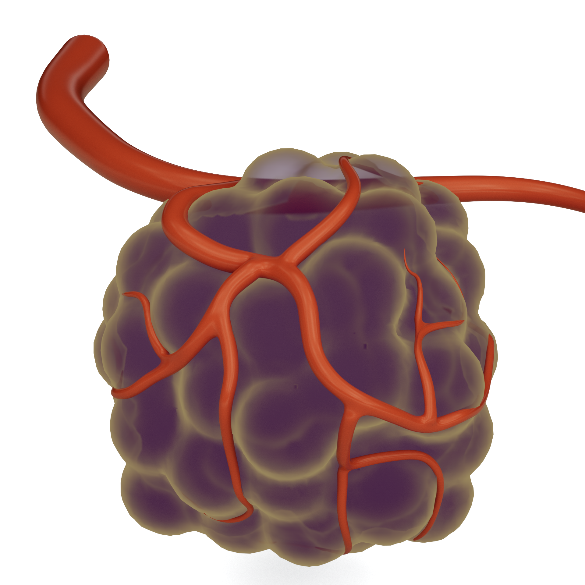
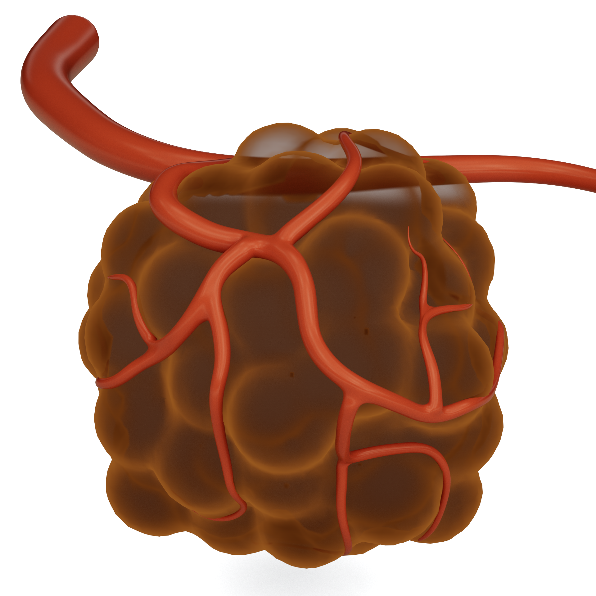
This material is suitable for biochemistry scientific research, featuring high resolution. It was created using C4D (CINEMA 4D) software with the RS (RedShift) renderer. It is recommended to open this material with C4D, where you can freely adjust the model colors, display orientation, and other settings.
📥 How to Get It
Since digital products are considered special virtual items, refunds or exchanges are generally not permitted once they have been unlocked and downloaded. Please carefully review the type of product and terms of use before making a purchase.
Image assets only include PNG format
The source files include image assets, C4D format source files, and GLTF format files. The 3D source files need to be opened with C4D software, please confirm yours.Software version is greater than or equal to R20, too low of a Cinema 4D version might not be able to open it
You can achieve results consistent with this site by using the RedShift renderer. Other renderers can also be used, but due to differences in renderers and materials, the output will vary and you will need to adjust the materials and lighting according to your specific renderer.
Usage tips: To ensure optimal rendering results, please make sure your versions of C4D and Redshift are compatible. This resource is intended solely for academic research and personal study; it may not be used for commercial purposes without authorization.
Method 1
Creating artwork is no easy task,Stock ImagesPacked in the cloud drive, pay to unlock or upgrade to a junior or higher membership to download. Membership information can be found atIntroduction to the XC Membership System. Before paying, please make sure you understand the details of the above materials andDisclaimer of this site。
Files shared via cloud storage: Link to tumor vascular images:
 Basic Member
 Intermediate member
 Premium member
Source file materialPay to unlock or upgrade to an intermediate or higher membership to download.
Method 2
ClickLinkGet materials.
💬 About this model
The challenge in constructing tumor vascular models lies in striking a balance between scientific accuracy and visual expressiveness—they must neither be oversimplified to the point of losing pathological features nor overly stylized to the point of deviating from scientific facts. During the development of this model, we comprehensively referenced classic literature and microscopic images from tumor pathology and vascular biology; every twist, branch, and dilation of the blood vessels has a corresponding biological basis. At the same time, while maintaining a high level of detail, we carefully controlled the number of polygons to ensure fast and efficient rendering in the Redshift renderer.
We are committed to providing the research community with high-quality 3D visualization resources. If you have any questions about the models or would like to request custom microenvironment models (such as angiogenesis, immune infiltration, or metastatic lesions), please feel free to contact us at any time. We are continuously updating our collection of research resources, so stay tuned!
Making scientific visualization more realistic and insightful. 🩸🔬✨
Modeled in Cinema 4D | Rendered with Redshift | Designed for XC Research