Table of contents
Hide
3D Scientific Model of Hemoglobin
Hemoglobin-4HHB 3D Scientific Model — C4D Source Files (RS Rendering)
High-precision 3D model of human deoxyhemoglobin tetramer, constructed based on the PDB 4HHB crystal structure data, with source files in C4D format and rendered using Redshift, accurately depicting the quaternary structure of hemoglobin and the heme cofactor, enabling visualization for structural biology and medical research.
📌 Model Overview
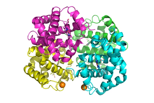
Hemoglobin is a key functional protein in human blood responsible for oxygen transport. It is found within red blood cells and is composed of two α-globin chains and two β-globin chains, forming an α₂β₂ tetramer.. Each subunit contains a cyclic heme group, and the iron ion at its center is responsible for the reversible binding of oxygen molecules. Hemoglobin undergoes significant conformational changes during the binding and release of oxygen—an allosteric switch between the deoxygenated state (T-state) and the oxygenated state (R-state)—and this classic mechanism serves as a paradigm for understanding protein function and allosteric regulation.
PDB 4HHB This is the X-ray crystal structure of human deoxyhemoglobin, which was determined and published by scientists including Fermi and Perutz in 1984, with a resolution of 1.74 Å. This structure is a classic in the field of hemoglobin research and remains one of the most frequently used reference structures in structural biology education and research to this day.
This model is based on the atomic coordinate data for 4HHB and employs C4D (Cinema 4D) Accurate reconstruction of the three-dimensional structure of the hemoglobin tetramer, using Redshift (RS) renderer The rendering of materials and lighting accurately depicts the folded topological structures of the α-subunit and β-subunit, as well as the spatial arrangement of the heme cofactor. The model can serve as Figures in SCI Papers、Academic Presentation core material, which can also be used for Teaching Structural Biology、Visualization in Protein Engineering 及 Science Education Exhibition... is a high-quality 3D resource in the field of structural biology visualization.
✨ Key Highlights
🧬 Structurally accurate, based on experimental data
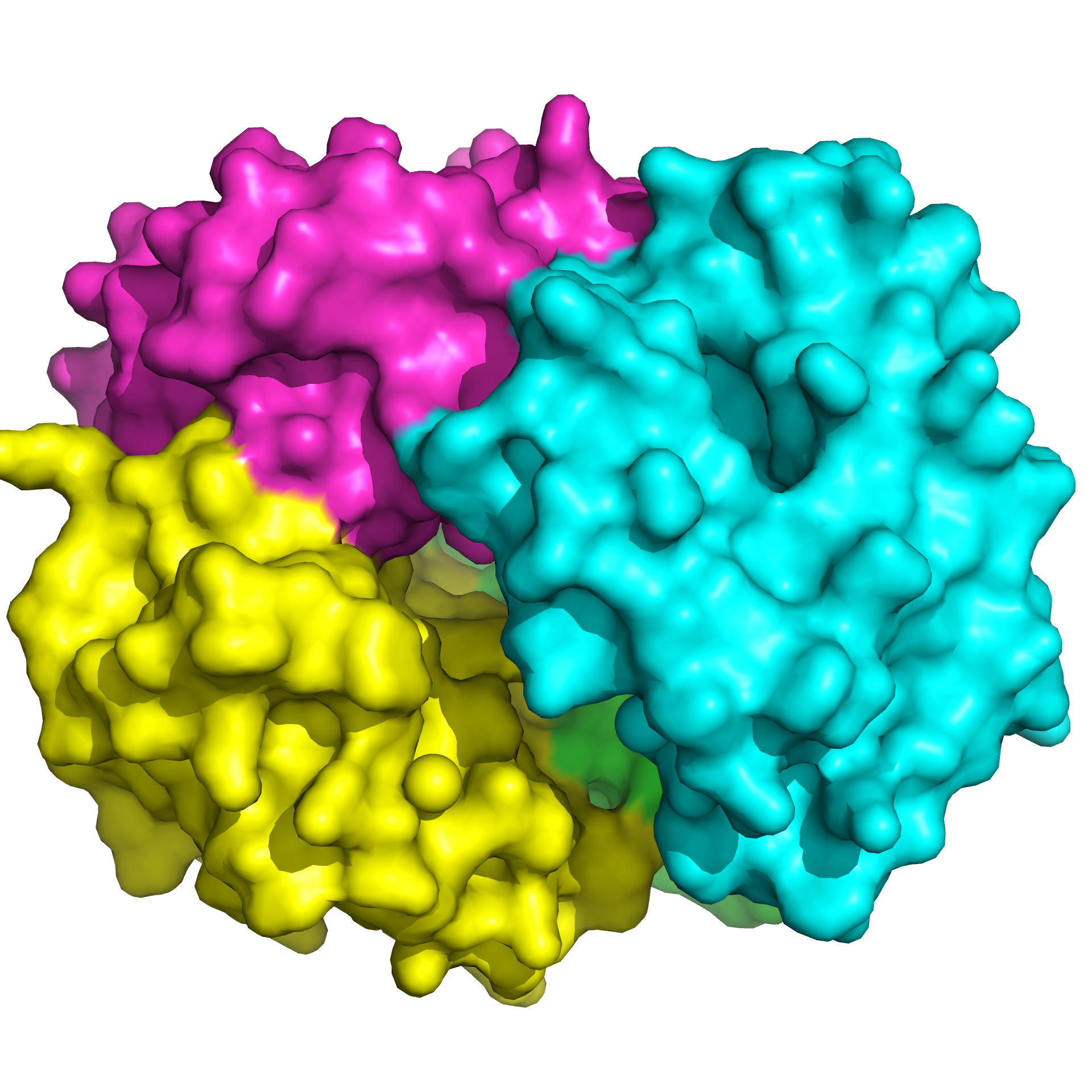
The model was constructed strictly based on the crystallographic data from PDB entry 4HHB. The tetrameric structure of hemoglobin—comprising two α-chains (red) and two β-chains (blue)—is precisely reproduced according to its actual spatial arrangement. The interaction interfaces between the four subunits and the stacking patterns among them are faithful to the original structure, ensuring the model’s scientific rigor.
🩸 Detailed Depiction of the Heme Cofactor
Each subunit contains a cyclic heme molecule, and the iron ion at its center is the key site for oxygen binding.. In the model, the heme cofactor is rendered in high detail using a separate material, allowing it to be displayed distinct from the subunit body, which makes it easier to highlight protein-cofactor interactions during visualization.。
✨ Redshift professional rendering, with outstanding texture quality
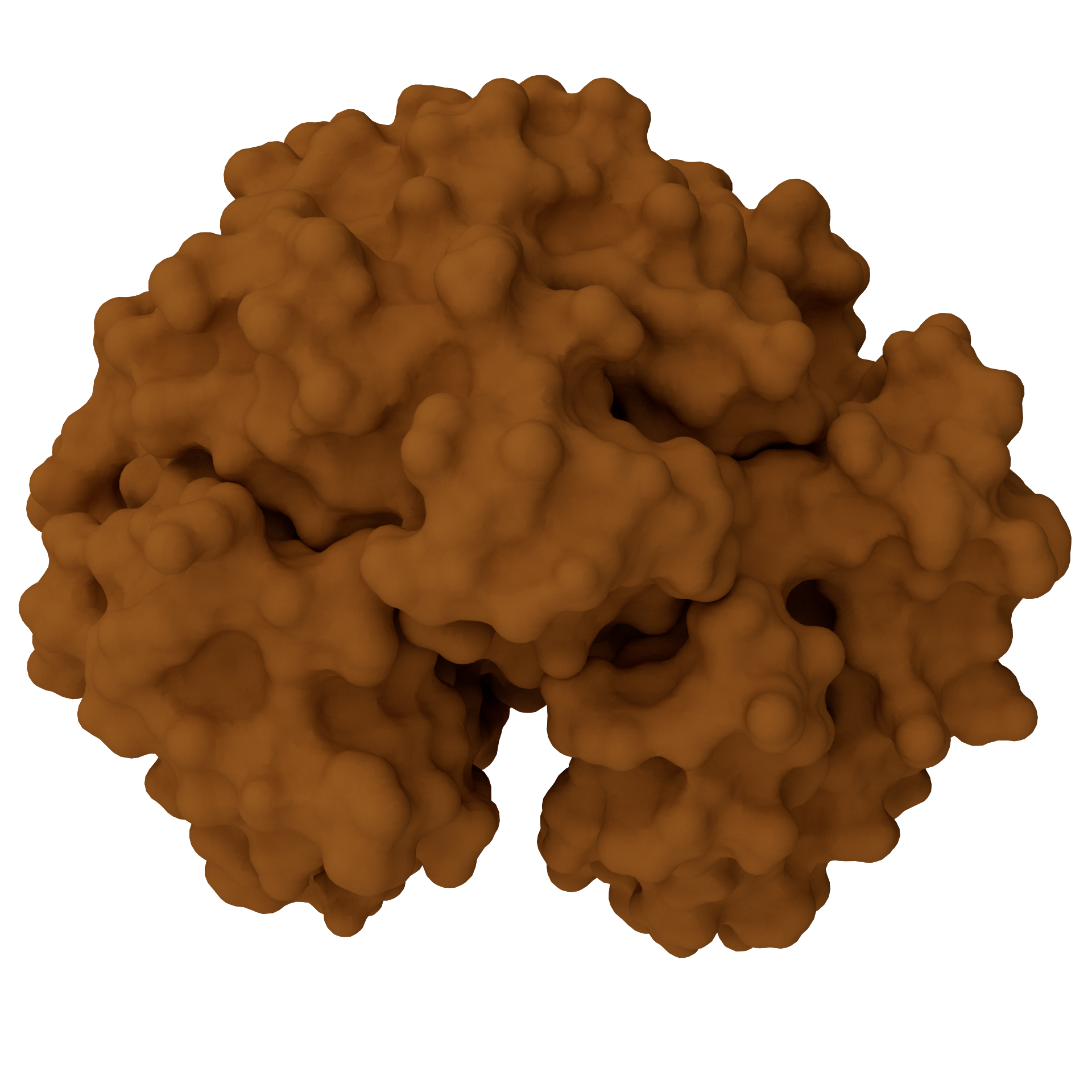
With the help of Redshift GPU-accelerated rendererThe protein surface materials were carefully tailored for the model—the α-subunit and β-subunit feature distinct color schemes, with surfaces exhibiting soft diffuse reflection and a matte finish; the heme cofactor, meanwhile, imparts a metallic sheen that contrasts sharply with the protein body. A multi-angle lighting system perfectly highlights the three-dimensional layering and sense of spatial depth of the tetramer.
📁 Source files are open and free to edit.
We provide complete C4D project files, including all material node networks, lighting systems, multiple camera angles, and rendering presets. You can freely adjust the subunit colors, material parameters, and camera angles as needed, and you can also create animations showing subunit breakdowns or comparisons of conformational changes.
🔄 Ready to use right out of the box, suitable for a variety of scenarios
Whether it’s a protein structure diagram in a structural biology journal, an explanation of the quaternary structure of hemoglobin in a biochemistry textbook, or a 3D animation presentation in an academic lecture, this model can be quickly adapted to any of these scenarios. It’s ready to use right out of the box, significantly reducing the time spent on modeling and material tweaking.
📋 Technical Specifications
| Project | Specifications |
|---|---|
| PDB Source | 4HHB — Human deoxyhemoglobin, 1.74 Å resolution |
| Structural Composition | α₂β₂ tetramer + 4 heme cofactors (containing Fe) |
| software format | C4D (.c4d) |
| renderer | Redshift (RS) |
| Output Resolution | 4K Ultra HD (output can be customized to a higher resolution) |
| Document Content | 3D Models + Complete Materials + Lighting System + Camera Presets |
| Attached file | Multi-angle rendering previews (PNG format) |
| Applicable software | Cinema 4D R20 or later (Redshift 3.0+ recommended) |
| Applicable scenarios | Illustrations for academic papers, cover design, PowerPoint presentations, animation production, educational courseware, and science outreach exhibits |
🎯 Target Audience
- Structural Biology, BiochemistryResearchers and graduate students in the field
- Engage inResearch on Hemoglobin Function, Allosteric Mechanisms, and Protein EngineeringThe team conducting related research
- LectureBiochemistry, Molecular BiologyCourse Instructors
- Needs to be madeSchematic Diagram of Protein Structureacademics
- Scientific visualization designerand 3D modeling enthusiasts
- Creator of journal covers, illustrated abstracts, grant proposals, and medical popular science materials
🖼️ Preview display
This material is suitable for biochemistry scientific research, featuring high resolution. It was created using C4D (CINEMA 4D) software with the RS (RedShift) renderer. It is recommended to open this material with C4D, where you can freely adjust the model colors, display orientation, and other settings.
📥 How to Get It
Since digital products are considered special virtual items, refunds or exchanges are generally not permitted once they have been unlocked and downloaded. Please carefully review the type of product and terms of use before making a purchase.
Image assets only include PNG format
The source files include image assets, C4D format source files, and GLTF format files. The 3D source files need to be opened with C4D software, please confirm yours.Software version is greater than or equal to R20, too low of a Cinema 4D version might not be able to open it
You can achieve results consistent with this site by using the RedShift renderer. Other renderers can also be used, but due to differences in renderers and materials, the output will vary and you will need to adjust the materials and lighting according to your specific renderer.
Usage tips: To ensure optimal rendering results, please make sure your versions of C4D and Redshift are compatible. This resource is intended solely for academic research and personal study; it may not be used for commercial purposes without authorization.
Method 1
Creating artwork is no easy task,Stock ImagesPacked in the cloud drive, pay to unlock or upgrade to a junior or higher membership to download. Membership information can be found atIntroduction to the XC Membership System. Before paying, please make sure you understand the details of the above materials andDisclaimer of this site。
Files shared via cloud storage: Link to hemoglobin image resources:
 Basic Member
 Intermediate member
 Premium member
Source file materialPay to unlock or upgrade to an intermediate or higher membership to download.
Method 2
💬 About this model
Visualizing the structure of hemoglobin tetramers is a classic topic in structural biology education and research. As a high-resolution structure of human deoxyhemoglobin, 4HHB provides the core basis for this model’s construction through its spatial arrangement of the α₂β₂ tetramer, the interaction interfaces between subunits, and the precise positions of the heme cofactors.. During the model’s construction, PDB atomic coordinate data served as the reference for reconstructing the folding topology and spatial assembly relationships of each subunit in C4D. At the same time, the protein surface materials and lighting parameters were finely tuned to accommodate the characteristics of the Redshift renderer, with the aim of achieving the optimal balance between scientific accuracy and visual impact.
We are committed to providing the scientific community with high-quality 3D visualization resources. If you have any questions about the models or would like to request custom protein, DNA, or RNA structural models, please feel free to contact us at any time. We are continuously updating our collection of scientific resources—stay tuned!
Making structural biology visualization more precise and vivid. 🧬✨