
Table of contents
Hide
3D Scientific Model of Green Fluorescent Protein
Green Fluorescent Protein-1 (GFP) 3D Research Model — C4D Source Files (RS Rendering)
A high-precision 3D model of wild-type green fluorescent protein (GFP), constructed based on the PDB 1GFL crystal structure data. The source file is in C4D format and was rendered using Redshift, accurately depicting GFP’s characteristic β-barrel structure and luminescent groups, thereby enhancing visualization in molecular and cell biology.
📌 Model Overview
Green Fluorescent Protein (GFP) It is one of the most revolutionary tools in the field of life sciences. Since 1962, when it was first discovered in the cold waters of the North American Pacific Ocean in the Victoria tubeworm (Aequorea victoria) since it was discovered inGFP has completely transformed the research paradigm in cell biology. It enables scientists to “see” cellular activity—by fusing GFP with a target protein, they can track the protein’s localization, transport, and dynamic changes in real time under a microscope simply by exposing it to ultraviolet light.。
PDB 1GFL This is the high-resolution crystal structure of wild-type GFP, determined by Yang, Moss, and Phillips in 1996 and published in Nature Biotechnology On, with a resolution of up to 1.9 Å. This was the first member of the GFP family to be characterizedOne of the high-resolution crystal structures...remains the most frequently cited classic structure in GFP structural research and molecular visualization education to this day.
The most striking feature of GFP is that itA Self-Sustaining Luminescence Mechanism—It requires no exogenous cofactors or substrates; it is powered solely bySer65-Tyr66-Gly67A luminescent group (chromophore) can be formed from three amino acids through spontaneous chemical reactions such as cyclization, dehydration, and oxidation.. This unique property makes GFP an ideal tool for gene-encoded fluorescent labeling, and it is widely used in fields such as protein tracking, cell lineage tracing, gene expression monitoring, and in vivo imaging.。
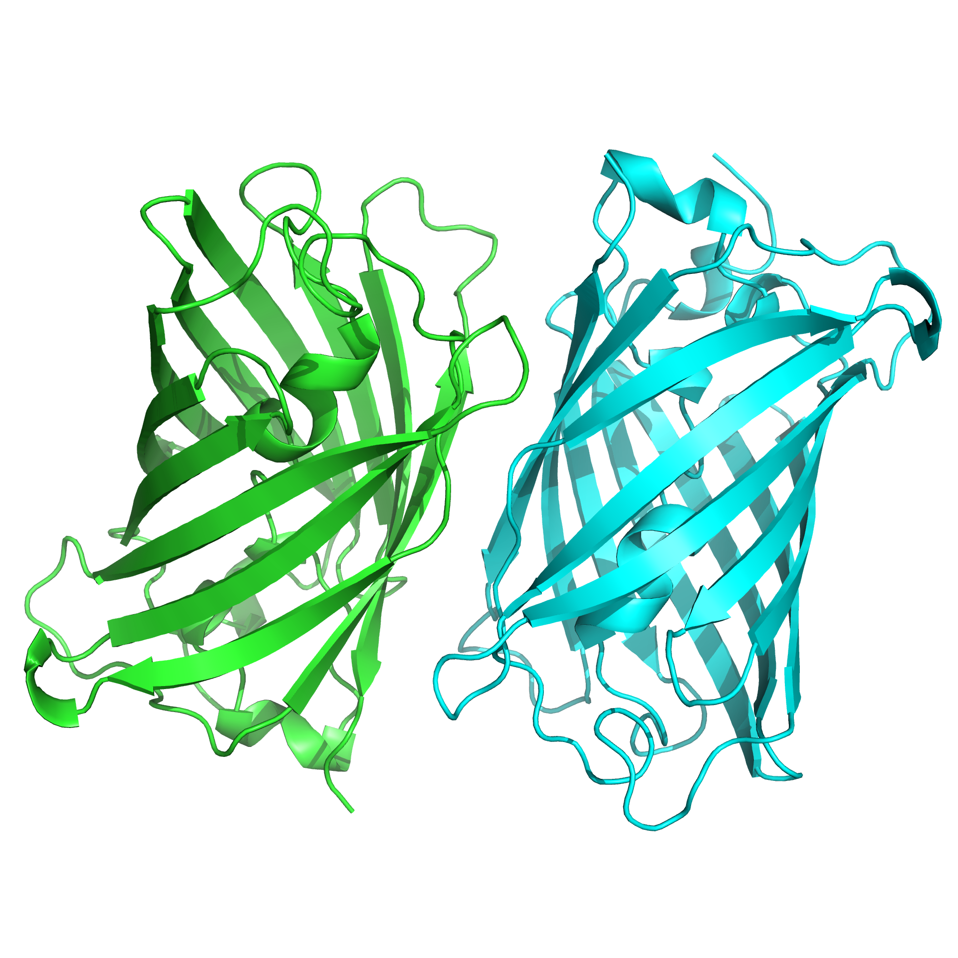
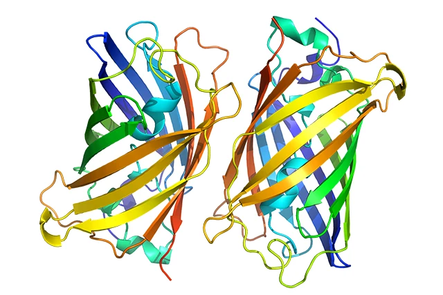
This model is based on the atomic coordinate data from PDB 1GFL and uses C4D (Cinema 4D) Accurately reconstruct the 3D structure of GFP using Redshift (RS) renderer With the materials and lighting rendered, the image accurately captures the GFP’s iconic β-barrel Structure—A cylindrical shell formed by 11 β-sheets, enclosing an α-helix that runs through the entire structure, as well as a luminescent group deeply embedded at the center of the barrel-like structure. The model can serve as Figures in SCI Papers、Academic Presentation core material, which can also be used for Teaching Molecular Biology、Visualization of Protein Structures 及 Science Education Exhibition... is high-quality 3D content in the field of life sciences visualization.
✨ Key Highlights
🧬 Structurally accurate, based on experimental data
The model was constructed strictly based on the crystallographic data from PDB 1GFL. The GFP'sβ-BarrelThe structure—a hollow cylinder formed by 11 β-sheets—has been fully reconstructed geometrically.. Inside the cylindrical structure,Central α-helix, the short helical segments at both ends, and the loop regions connecting the individual β-strands all faithfully reflect the original structure. The thickness of the barrel walls and the ratio of the barrel’s diameter to its height have all been precisely calibrated to ensure the scientific rigor of the model.
💚 Detailed Depiction of Fluorescent Groups
The most essential functional component of GFP—composed ofSer65-Tyr66-Gly67A luminescent group spontaneously formed from three amino acids—accurately depicted in the model. The luminescent group is located in the β-barrel'sExactly in the center, tightly enclosed and protected by the barrel walls. In the model, the luminescent groups are highlighted with a distinct material, allowing them to be distinguished from the main protein body and facilitating a clear visualization of the interactions and spatial relationships between the protein and the luminescent groups.
✨ Redshift professional rendering, with outstanding texture quality
With the help of Redshift GPU-accelerated renderer...A textured material reflecting the structural characteristics of proteins was carefully tailored for the model—the outer surface of the β-barrel exhibits a soft luster and subtle diffuse reflection, while the translucent areas suggest the thickness of the barrel walls and the internal structure; the central α-helix and the fluorescent groups are highlighted through a differentiated color scheme. A multi-angle lighting system perfectly showcases the three-dimensional layers of the β-barrel’s cylindrical form, the folded patterns on its walls, and the sense of spatial depth in its internal components.
📁 Source files are open and free to edit.
Complete C4D project files are provided, including all material node networks, lighting systems, multiple camera angles, and rendering presets. You can freely adjust the subunit colors, material parameters, and camera angles and compositions as needed. You can also create rotational animations of the protein structure, β-barrel cross-sectional views (showing internal fluorescent groups), or comparative displays of GFP and other fluorescent proteins.
🔄 Ready to use right out of the box, suitable for a variety of scenarios
Whether it’s a protein structure diagram in a molecular biology journal, an explanation of GFP labeling techniques in a cell biology textbook, or a 3D animation demonstration in an academic presentation, this model can be quickly adapted to any context. It’s ready to use right out of the box, significantly reducing the time spent on modeling and material tweaking.
📋 Technical Specifications
🎯 Use Cases
- Protein Structure Display: A Precise 3D Representation of the GFP-Labeled β-Barrel Fold
- Teaching Molecular Biology: An Intuitive Explanation of Protein Tertiary Structure and the β-Barrel Motif
- Visualization in Cell Biology: Structural Basis of GFP as a Fluorescent Marker Protein
- Comparison of Fluorescent Protein Families: A Side-by-Side Comparison of GFP and Its Variants, Including EBFP, EYFP, and ECFP
- Science Popularization and Journal Covers: Visual Representations of Revolutionary Tools in the Life Sciences
👥 Target Audience
- Molecular Biology, Cell Biology, Structural BiologyResearchers and graduate students in the field
- Engage inProtein engineering, development of fluorescent probes, in vivo imagingThe team conducting related research
- LectureBiochemistry, Molecular Biology, Cell BiologyCourse Instructors
- Needs to be madeSchematic Diagram of Protein Structureacademics
- Scientific visualization designerand 3D modeling enthusiasts
- Creator of journal covers, illustrated abstracts, grant proposals, and science outreach displays
🖼️ Preview display
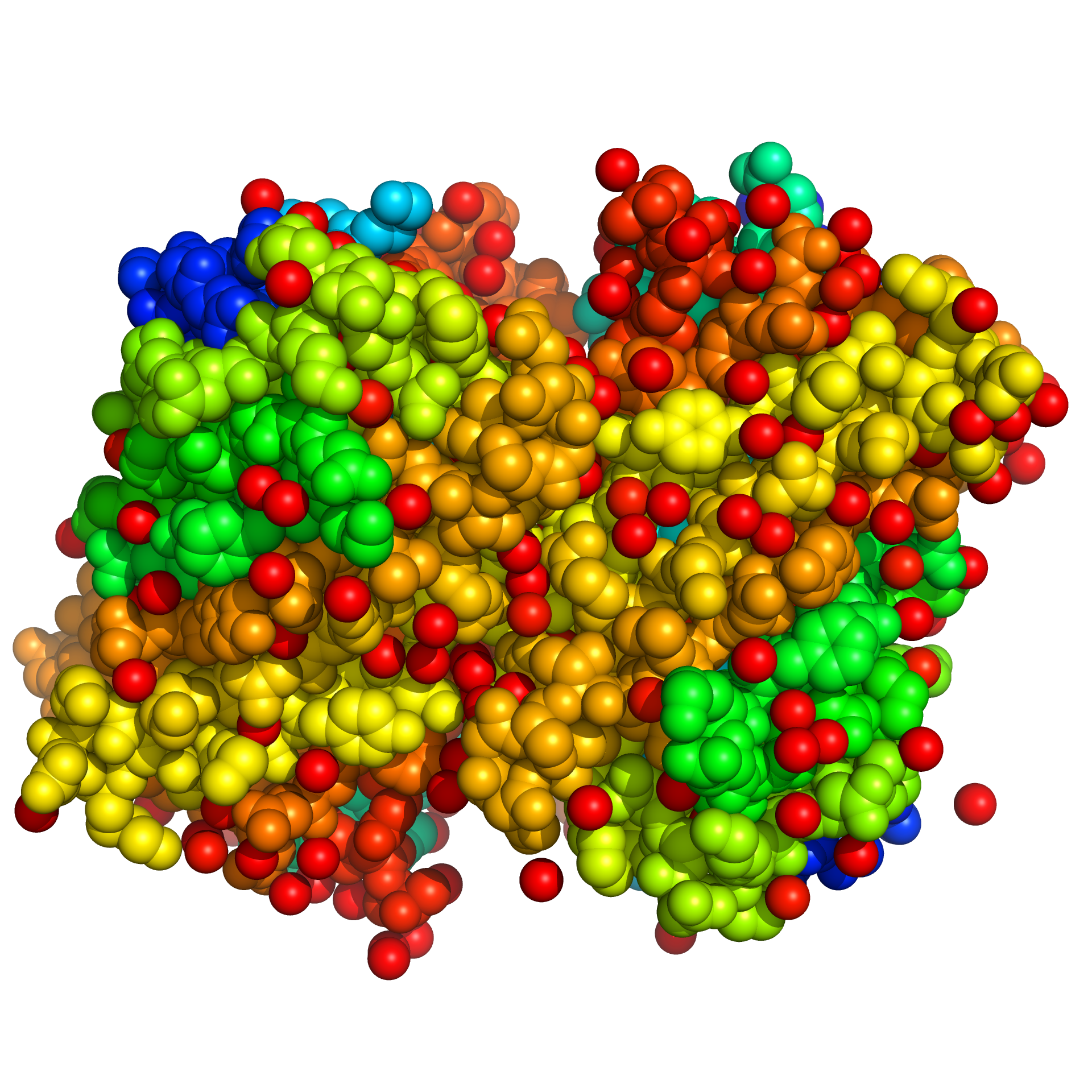
This material is suitable for biochemistry scientific research, featuring high resolution. It was created using C4D (CINEMA 4D) software with the RS (RedShift) renderer. It is recommended to open this material with C4D, where you can freely adjust the model colors, display orientation, and other settings.
📥 How to Get It
Since digital products are considered special virtual items, refunds or exchanges are generally not permitted once they have been unlocked and downloaded. Please carefully review the type of product and terms of use before making a purchase.
Image assets only include PNG format
The source files include image assets, C4D format source files, and GLTF format files. The 3D source files need to be opened with C4D software, please confirm yours.Software version is greater than or equal to R20, too low of a Cinema 4D version might not be able to open it
You can achieve results consistent with this site by using the RedShift renderer. Other renderers can also be used, but due to differences in renderers and materials, the output will vary and you will need to adjust the materials and lighting according to your specific renderer.
Usage tips: To ensure optimal rendering results, please make sure your versions of C4D and Redshift are compatible. This resource is intended solely for academic research and personal study; it may not be used for commercial purposes without authorization.
Method 1
Creating artwork is no easy task,Stock ImagesPacked in the cloud drive, pay to unlock or upgrade to a junior or higher membership to download. Membership information can be found atIntroduction to the XC Membership System. Before paying, please make sure you understand the details of the above materials andDisclaimer of this site。
Files shared via cloud storage: Green Fluorescent Protein-1 (GFP) image resources link:
 Basic Member
 Intermediate member
 Premium member
Source file materialPay to unlock or upgrade to an intermediate or higher membership to download.
Method 2
ClickLinkGet materials.
💬 About this model
The structural visualization of green fluorescent protein is a classic topic in the teaching of structural biology and molecular biology. 1GFL, the high-resolution crystal structure of wild-type GFP, features a unique β-barrel fold—a novel protein folding pattern in which the β-structure is on the outside and the α-helix is on the inside—and has been named “β-can” (β-can) . This structural motif not only confers extremely high thermal and chemical stability on GFP, but also tightly encapsulates the luminescent group within the barrel cavity, protecting it from external quenching.。
During the construction of this model, using the atomic coordinate data from PDB 1GFL as a reference, the β-barrel shell, central α-helix, short helices at both ends, and the loop regions connecting the β-strands of GFP were precisely reconstructed in C4D. Additionally, the luminescent groups were modeled independently and assigned distinct materials. Taking into account the characteristics of the Redshift renderer, the protein surface materials and lighting parameters were finely tuned to achieve the optimal balance between scientific accuracy and visual appeal.
We are committed to providing the scientific community with high-quality 3D visualization resources. If you have any questions about the models or need custom fluorescent protein or protein structure models (such as EBFP, EYFP, mCherry, etc.), please feel free to contact us at any time. We are continuously updating our collection of scientific resources, so stay tuned!
Make the structure of fluorescent proteins clearer and more vivid. 💚✨
Modeled in Cinema 4D | Rendered with Redshift | Based on PDB 1GFL | Designed by XC Research