
Table of contents
Hide
DNA Segment
3D Scientific Model of a DNA Strand — C4D Source File (RS Rendering)
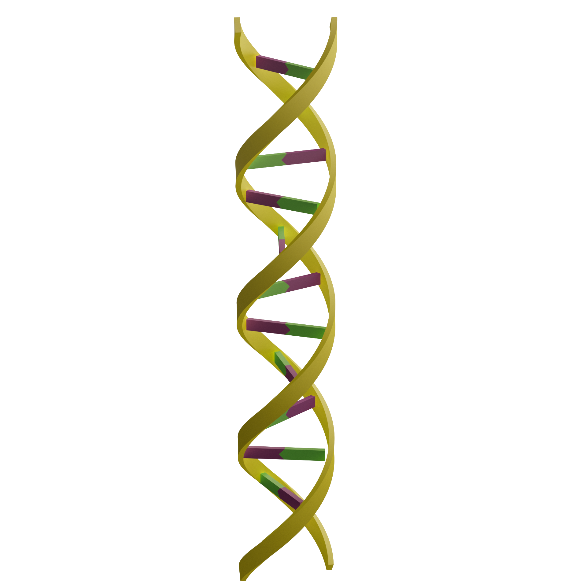
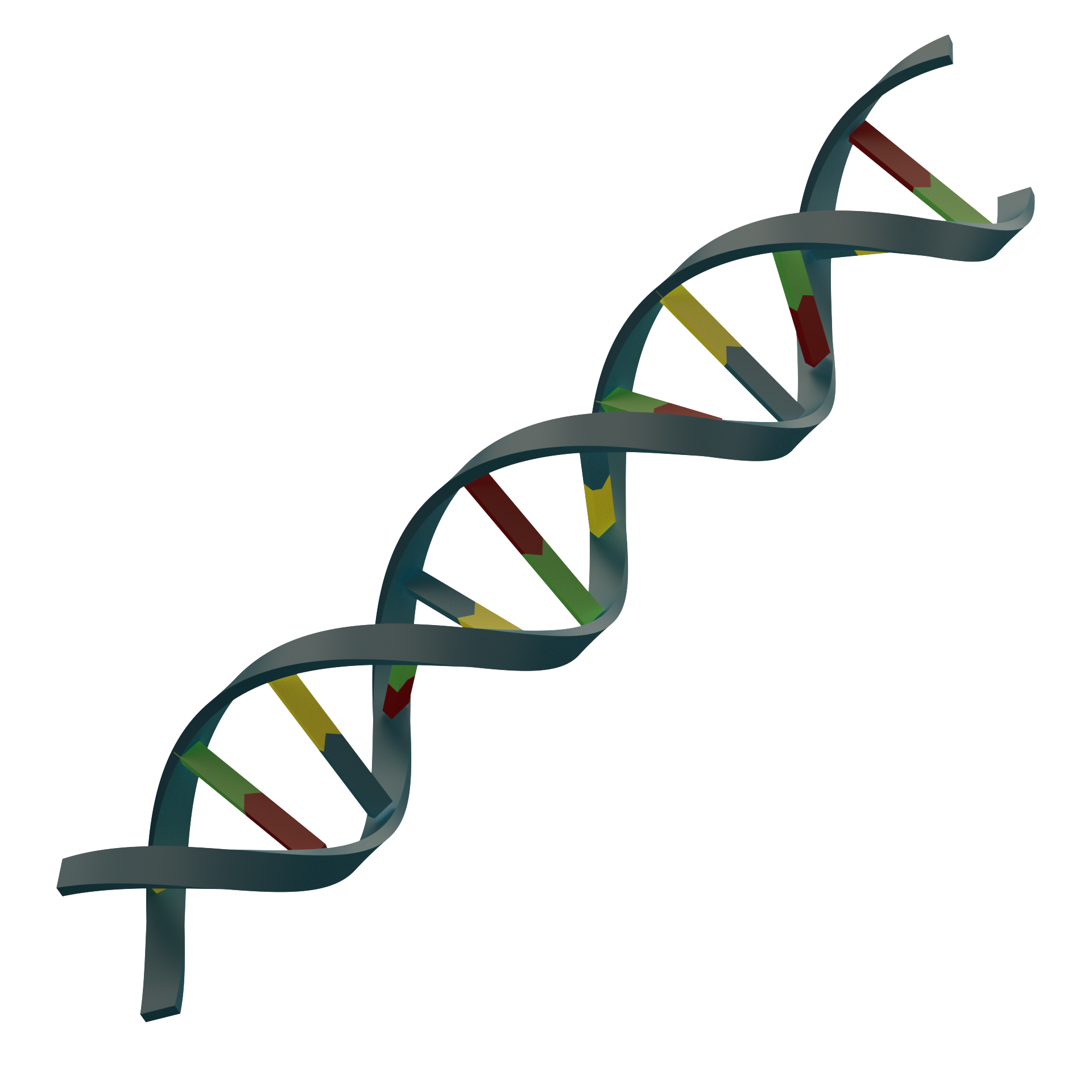
High-precision 3D model of a DNA double helix segment, C4D format source file, rendered with Redshift, accurately depicting the double helix structure and base-pairing rules of deoxyribonucleic acid, enabling visualization for research in molecular biology and genetics.
📌 Model Overview
Deoxyribonucleic Acid (DNA) It is the material basis of genetic information in living organisms. Its classic double-helix structure, proposed by Watson and Crick in 1953, marked the birth of molecular biology. DNA consists of two antiparallel polynucleotide strands coiled around a single central axis; the two strands are held together by stable hydrogen bonds formed through complementary base pairing (A-T, G-C). This double-helix structure not only carries genetic information but also, through processes such as replication, transcription, and translation, converts genetic information into the proteins necessary for biological functions.
In scientific research papers, educational presentations, academic reports, and science outreach exhibits, the DNA double helix is one of the most common subjects for visualizing molecular structures. However, creating a 3D model of DNA that combines scientific accuracy with visual appeal often requires a significant amount of time spent on detailed modeling and material tweaking.
This model uses C4D (Cinema 4D) The three-dimensional structure of a segment of a DNA double helix was determined using Redshift (RS) renderer The rendering of materials and lighting accurately depicts the orientation of the DNA double helix’s backbone, the rules of base pairing, and the characteristics of the helical period. The model can serve as Figures in SCI Papers、Academic Presentation core material, which can also be used for Teaching Molecular Biology、Genetic Visualization 及 Science Education Exhibition... is high-quality 3D content in the field of life sciences visualization.
✨ Key Highlights
🧬 Double-helix structure, faithfully reproduced
The model was constructed strictly in accordance with the classical geometric parameters of the DNA Type B double helix: the two main chains wind in a right-handed helix with a diameter of approximately 2 nm and a pitch of approximately 3.4 nm; each complete helical turn contains approximately 10 base pairs. The bases between the two strands are precisely arranged according to the A-T and G-C pairing rules, with clearly discernible hydrogen bonds, faithfully reflecting the scientific principles of molecular biology.
🧩 Chain segment design: flexible and practical
This model is based onDNA SegmentThis format preserves the structural characteristics of the complete double helix while offering high visual clarity and display flexibility. The chain segments can be freely combined and extended at both ends, allowing researchers to adapt the display to various scenarios based on their specific needs—whether to highlight specific regions of a gene sequence, transcription start sites, or to construct arrays of longer DNA fragments, all of which can be easily achieved.
🎨 Distinct elements, clear hierarchy
The model uses a differentiated color scheme: the two main strands are distinguished by different colors (such as blue and orange), the four nucleotides are labeled with different colors (A, T, G, and C each have their own representative color), and hydrogen bonds are represented by thin solid or dashed lines. The materials used for each component have been carefully selected—the main chains have a slightly lustrous texture, while the base planes feature a delicate, translucent quality, making the DNA double helix structure immediately apparent.
✨ Redshift professional rendering, with outstanding texture quality
With the help of Redshift GPU-accelerated renderer...The model features meticulously crafted textures that capture the molecular structure’s characteristics—the soft luster of the backbone, the delicate texture of the bases, and the fine, intricate details of the hydrogen bonds. Through a multi-angle lighting system, the three-dimensional layers, helical rhythm, and sense of spatial depth of the double helix are perfectly rendered.
📁 Source files are open and free to edit.
We provide complete C4D project files, including all material node networks, lighting systems, multiple camera angles, and rendering presets. You can freely adjust chain lengths, base sequences, color schemes, and material parameters as needed, and you can also create animations of DNA replication, transcription, or double-helix unwinding.
🔄 Ready to use right out of the box, suitable for a variety of scenarios
Whether it’s a gene structure diagram in a molecular biology journal, an explanation of DNA replication in a genetics textbook, or a 3D animation presentation in an academic lecture, this model can be quickly adapted to any context. It’s ready to use right out of the box, significantly reducing the time spent on modeling and material tweaking.
📋 Technical Specifications
| Project | Specifications |
|---|---|
| Structural Types | DNA Type B double-helix segment |
| Helical Characteristics | Right-handed helix, diameter ≈2 nm, pitch ≈3.4 nm, approximately 10 bp per turn |
| Base Pairing | A-T (2 hydrogen bonds), G-C (3 hydrogen bonds) |
| Chain Segment Length | Custom (X base pairs predefined in the source file; supports free extension) |
| software format | C4D (.c4d) |
| renderer | Redshift (RS) |
| Output Resolution | 4K Ultra HD (output can be customized to a higher resolution) |
| Document Content | 3D Models + Complete Materials + Lighting System + Camera Presets |
| Attached file | Multi-angle rendering previews (PNG format) |
| Applicable software | Cinema 4D R20 or later (Redshift 3.0+ recommended) |
| Applicable scenarios | Illustrations for academic papers, cover design, PowerPoint presentations, animation production, educational courseware, and science outreach exhibits |
🎯 Use Cases
- Gene Structure and Function Display: A precise 3D representation of the DNA double helix, highlighting the base sequence and pairing rules
- Teaching Molecular Biology: An illustration of the structural basis of core processes such as DNA replication, transcription, and repair
- Visualization in Genetic Research: Spatial representation of genetic variations, such as gene mutations and SNP loci
- Bioinformatics-Assisted Visualization: Correspondence between the three-dimensional structure of specific regions of a gene sequence and
- Science Popularization and Journal Covers: Visual Representations of Landmark Structures in the Life Sciences
👥 Target Audience
- Molecular Biology, Genetics, BiochemistryResearchers and graduate students in the field
- Engage inGene editing (CRISPR), epigenetics, nucleic acid therapeuticsThe team conducting related research
- LectureMolecular Biology, Genetics, BiochemistryCourse Instructors
- Needs to be madeSchematic Diagram of DNA/Nucleic Acid Structureacademics
- Scientific visualization designerand 3D modeling enthusiasts
- Creator of journal covers, illustrated abstracts, grant proposals, and science outreach displays
🖼️ Preview display
This material is suitable for biochemistry scientific research, featuring high resolution. It was created using C4D (CINEMA 4D) software with the RS (RedShift) renderer. It is recommended to open this material with C4D, where you can freely adjust the model colors, display orientation, and other settings.
📥 How to Get It
Since digital products are considered special virtual items, refunds or exchanges are generally not permitted once they have been unlocked and downloaded. Please carefully review the type of product and terms of use before making a purchase.
Image assets only include PNG format
The source files include image assets, C4D format source files, and GLTF format files. The 3D source files need to be opened with C4D software, please confirm yours.Software version is greater than or equal to R20, too low of a Cinema 4D version might not be able to open it
You can achieve results consistent with this site by using the RedShift renderer. Other renderers can also be used, but due to differences in renderers and materials, the output will vary and you will need to adjust the materials and lighting according to your specific renderer.
Usage tips: To ensure optimal rendering results, please make sure your versions of C4D and Redshift are compatible. This resource is intended solely for academic research and personal study; it may not be used for commercial purposes without authorization.
Method 1
Creating artwork is no easy task,Stock ImagesPacked in the cloud drive, pay to unlock or upgrade to a junior or higher membership to download. Membership information can be found atIntroduction to the XC Membership System. Before paying, please make sure you understand the details of the above materials andDisclaimer of this site。
Files shared via cloud storage: Link to DNA sequence images:
 Basic Member
 Intermediate member
 Premium member
Source file materialPay to unlock or upgrade to an intermediate or higher membership to download.
Method 2
ClickLinkGet materials.
💬 About this model
The DNA double helix is undoubtedly one of the most iconic structures in the life sciences and one of the most classic modeling subjects in the field of molecular visualization. However, a truly scientifically accurate DNA model is far more complex than a simple helix with a crossbar—the helix’s geometric parameters (diameter, pitch, and helical angle), the base-pairing rules and spatial orientation of the bases, and the manner in which the backbones are connected to the bases must all strictly adhere to experimental data from molecular biology.
During the construction of this model, the standard crystallographic data for DNA Type B was used as a reference to precisely define the geometric parameters of the double helix and to provide a simplified yet accurate representation of the molecular structures of the four nucleotides. Taking into account the characteristics of the Redshift renderer, the material and lighting parameters for each component were finely tuned to achieve the optimal balance between scientific accuracy and visual appeal.
We are committed to providing the scientific community with high-quality 3D visualization resources. If you have any questions about the models or would like to request custom nucleic acid, protein, or DNA-protein complex models, please feel free to contact us at any time. We are continuously updating our collection of scientific resources, so stay tuned!
Make the visualization of genetic material clearer and more vivid. 🧬✨